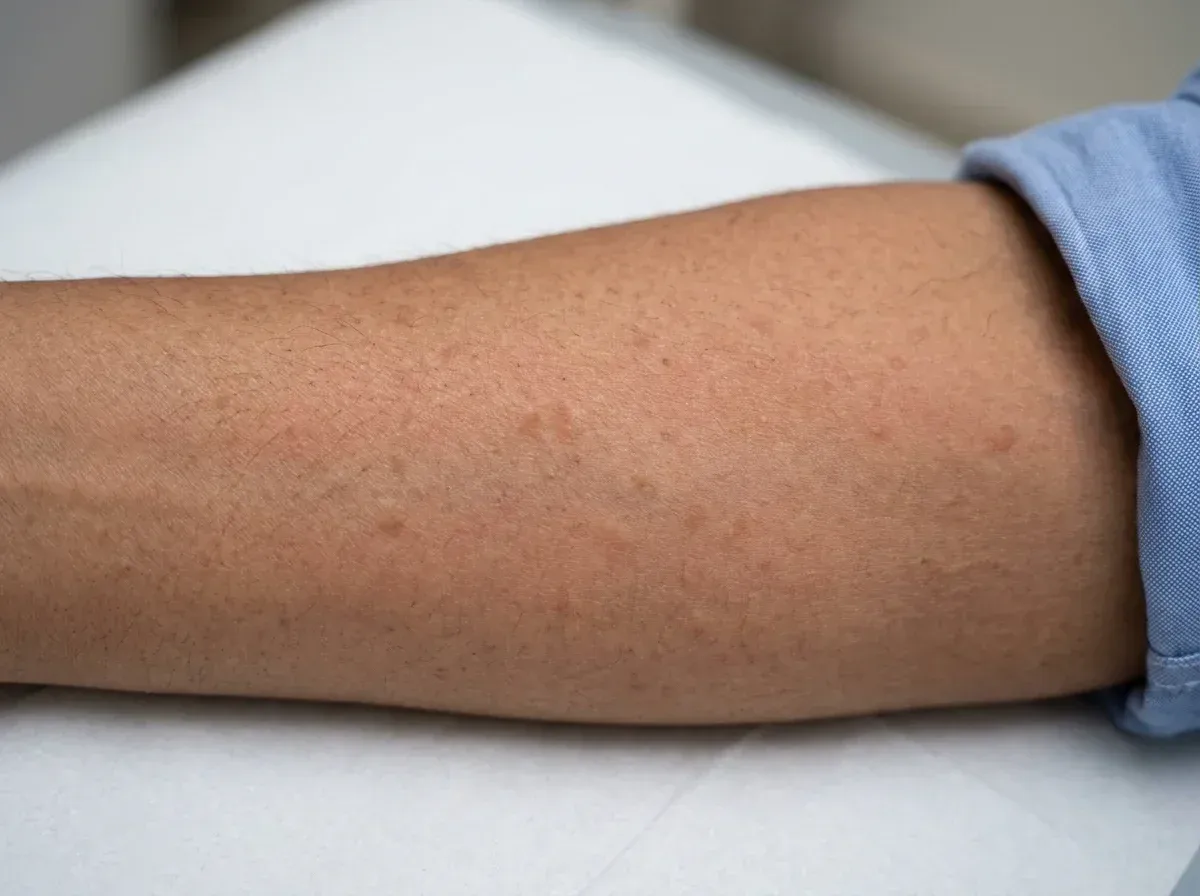
•Brownish-yellow spots on skin that itch and swell when rubbed (Darier's sign - pathognomonic)
•Recurrent flushing (sudden reddening of face and neck)
•Episodes of hives or severe itching
•Recurrent anaphylaxis without an obvious trigger
•Abdominal cramps, diarrhoea and nausea
•Bone pain or osteoporosis (in systemic disease)
•Fatigue and cognitive difficulties ('brain fog')
•Low blood pressure episodes and fainting
Causes & Triggers
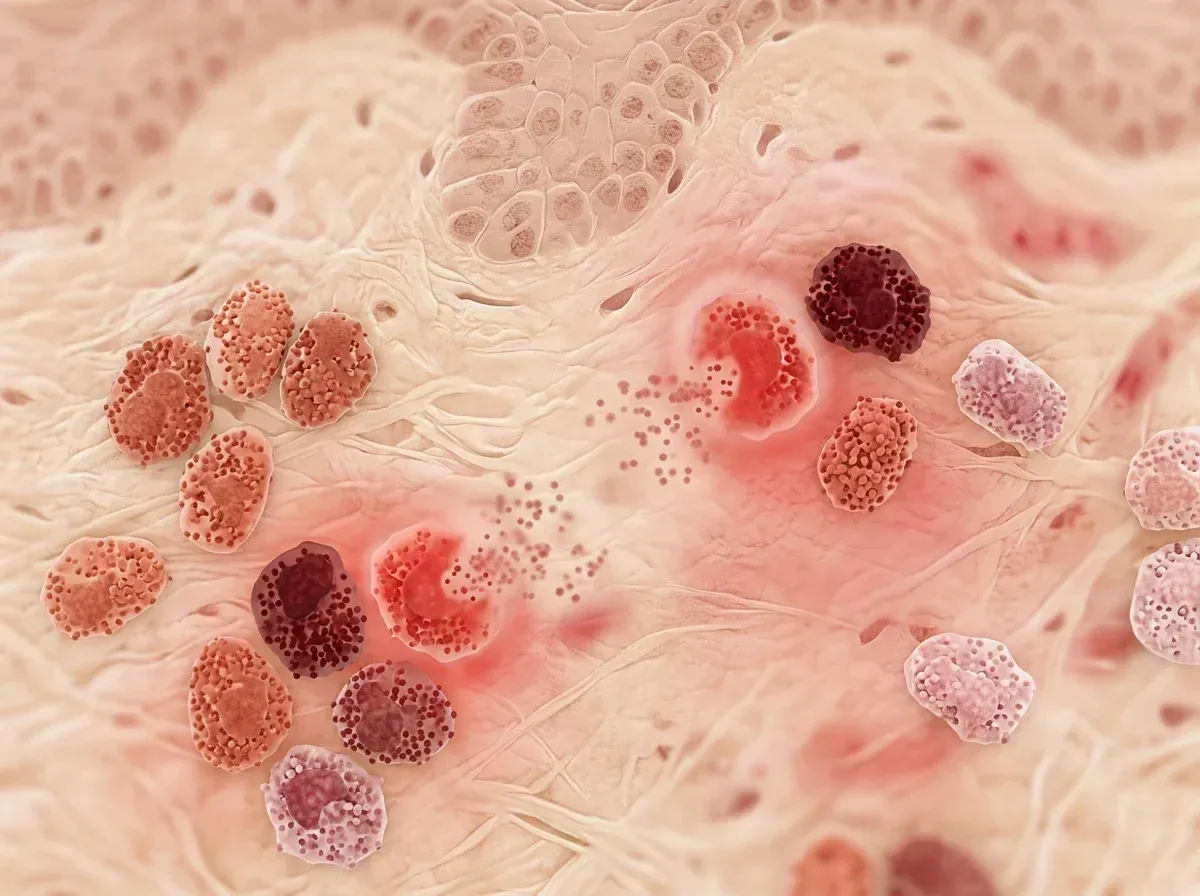
Mastocytosis is caused by a clonal proliferation of mast cells, most commonly driven by an acquired activating mutation in the KIT gene (D816V mutation in over 90% of systemic cases). This is not a conventional allergic disease - it is a clonal haematological disorder. In cutaneous mastocytosis (urticaria pigmentosa), the abnormal mast cells are confined to the skin; in systemic mastocytosis, they infiltrate the bone marrow, liver, spleen, and gastrointestinal tract. Environmental triggers that cause mast cell degranulation - and therefore symptom flares - include insect stings, NSAIDs (aspirin, ibuprofen), opioids (codeine), alcohol, extreme temperatures, mechanical friction, certain contrast dyes, and emotional stress. In Mumbai's climate of high heat and humidity, heat-triggered flushing and dermographism are common presenting complaints.
How We Test
Skin biopsy of pigmented lesions stained with Giemsa or toluidine blue reveals mast cell clusters - this is diagnostic for cutaneous mastocytosis. Serum tryptase level (basal) is the key systemic biomarker: a level above 20 ng/mL is a minor WHO diagnostic criterion for systemic mastocytosis. Bone marrow biopsy with KIT D816V mutation analysis (via PCR or next-generation sequencing) and immunohistochemistry (CD117+/CD25+ mast cells) is required to confirm systemic disease. Complete blood count, liver function tests, DEXA scan for bone density, and abdominal ultrasound complete the workup.
Management is aimed at symptom control, trigger avoidance, and in advanced cases, cytoreduction. All patients should avoid known triggers and carry an adrenaline auto-injector given the high risk of anaphylaxis. Symptom-directed therapy uses daily non-sedating H1 antihistamines (for itching and flushing), H2 blockers (for GI symptoms), and mast cell stabilisers (sodium cromoglycate, ketotifen). For aggressive systemic mastocytosis, targeted therapy with midostaurin (a KIT inhibitor) or avapritinib is available at select oncology centres in Mumbai. Bisphosphonates or denosumab address mastocytosis-associated osteoporosis.
Consult a specialist if you have recurring unexplained flushing, anaphylaxis without a clear trigger, or brownish itchy skin spots - especially if rubbing the spot causes a raised weal.
Frequently Asked Questions
Mastocytosis involves the same cells (mast cells) that drive allergic reactions, but the cells accumulate abnormally due to a gene mutation (KIT D816V) and release chemicals spontaneously - without requiring an allergen trigger. It is classified as a myeloproliferative neoplasm rather than an allergic disease, though it is best managed by an allergist-immunologist with expertise in mast cell disorders.
Darier's sign refers to the appearance of a raised, itchy weal (like an urticarial plaque) when a brownish skin lesion in mastocytosis is firmly stroked or rubbed. This occurs because rubbing mechanically degranulates the densely packed mast cells beneath the skin. A positive Darier's sign is a hallmark clinical finding that strongly supports the diagnosis of cutaneous mastocytosis.
Cutaneous mastocytosis in children often resolves spontaneously by puberty, so most paediatric patients experience a natural cure. In adults, systemic mastocytosis is generally a chronic condition managed rather than cured, though indolent forms have a normal life expectancy with appropriate symptom management. Aggressive variants are rare but require oncology-led therapy with KIT-inhibitor drugs.
Patients with mastocytosis have an abnormally large burden of mast cells, meaning that any trigger capable of causing mast cell degranulation can precipitate severe and sudden anaphylaxis, sometimes with no identifiable external allergen. An adrenaline auto-injector is a life-saving precaution that all patients with systemic mastocytosis - and some with extensive cutaneous disease - should carry at all times.
NSAIDs (aspirin, ibuprofen, diclofenac), opioid analgesics (codeine, morphine), some general anaesthetic agents, and iodinated radiocontrast media can provoke mast cell degranulation and severe reactions in mastocytosis patients. Patients should inform every doctor, dentist, and radiologist of their diagnosis, and a pre-medication protocol (antihistamines and steroids) is typically given before procedures or surgeries.
Take the next step
Living with mastocytosis? Let’s find your trigger.